
Kōkua kēia ʻatikala iā ʻoe e hoʻomaopopo maikaʻi i nā microscope ʻoki niho
ʻO ka microscope ʻoki niho, ma ke ʻano he "aniani hoʻonui nui" ma ke kahua o ka lāʻau lapaʻau waha, he mea hana pololei i hoʻohana kūikawā ʻia no ke ʻoki niho a me ka hōʻailona. Hōʻike maopopo ia i nā ʻano maalea i loko o ka lua waha i nā kauka ma o kahi moʻo o nā kūkulu paʻakikī a nani hoʻi, e hāʻawi ana i ka hiki ke mālama pololei.
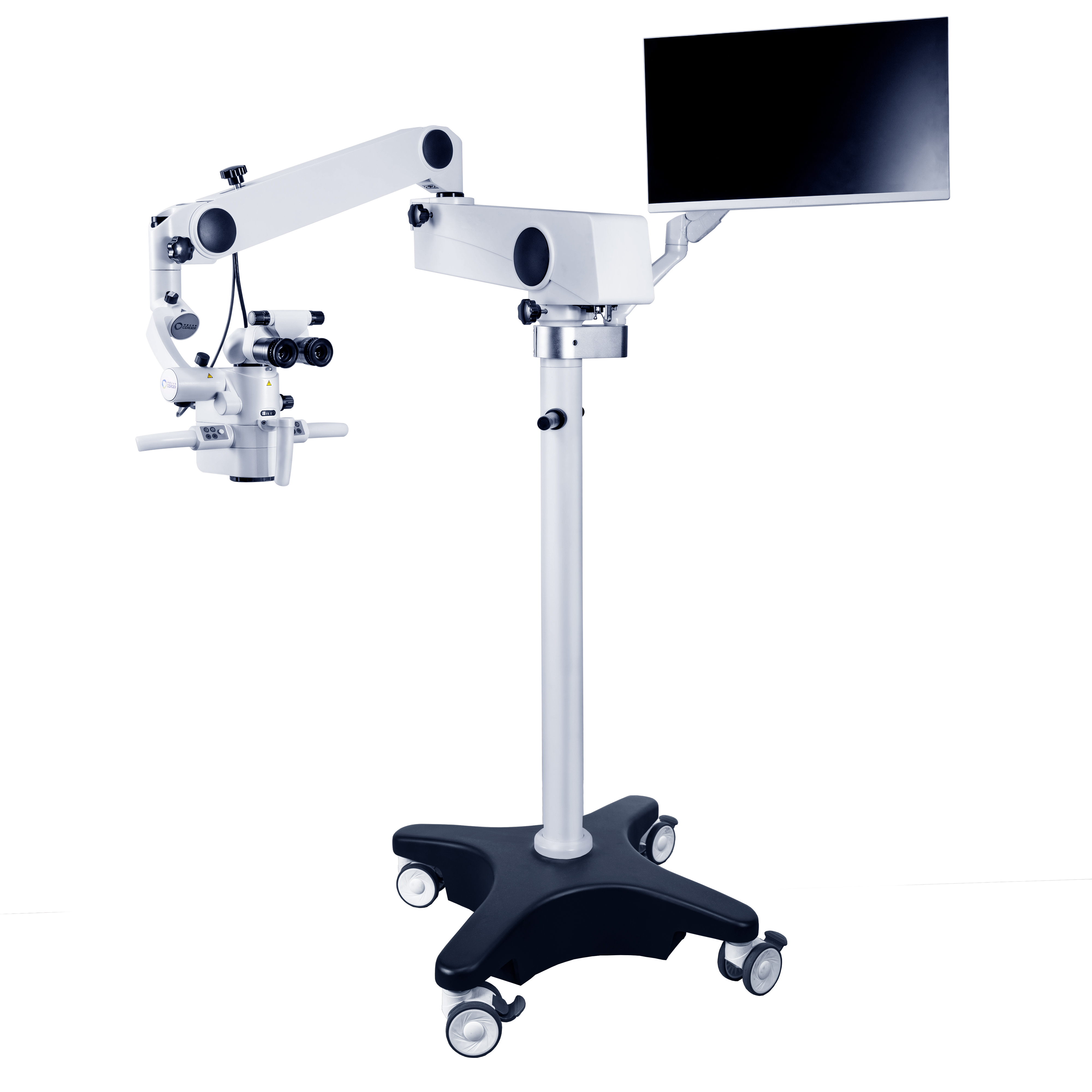
Mai kahi kuanaʻike hoʻonohonoho,nā microscope ʻoki nihoʻo ia hoʻi nā ʻāpana koʻikoʻi penei:
ʻŌnaehana hoʻonui optical:ʻO kēia kekahi o nā ʻāpana koʻikoʻi o kahimicroscope, e like me ke aniani o kahi kāmela, ka mea e hoʻoholo ai i ka hoʻonui ʻana a me ka moakāka o ke kiʻi. ʻO ka hoʻonui ʻana onā microscope ʻoki niho houʻO ka maʻamau, hiki ke hoʻololi ʻia ma waena o 4-40 mau manawa, a hiki i nā kauka ke hoʻololi maʻalahi i ka hoʻonui ʻana e like me nā pono o ke ʻoki kino, e like me ka hoʻoponopono ʻana i ka lōʻihi o ke kāmela. ʻO ka hoʻonui haʻahaʻa (4-8 mau manawa) he kūpono no ka nānā ʻana i kahi kahua ʻoki kino nui, e like me ka nānā ʻana i ke kūlana holoʻokoʻa o ka wahi ʻoki kino i ka wā o ke ʻoki waha; Hoʻokō ka hoʻonui waena (8-14 mau manawa) i nā pono o ka hapa nui o nā ʻoki niho maʻamau, e like me ka mālama ʻana i ke aʻa canal, ke ʻoki periodontal, a pēlā aku; ʻO ka hoʻonui kiʻekiʻe (14-40 mau manawa) e ʻae i nā kauka e ʻike i nā ʻano maalea loa, e like me nā lālā aʻa canal a me nā tubules dentinal i loko o nā niho, e hāʻawi ana i ke kākoʻo ikaika no nā hana maikaʻi.
ʻŌnaehana kukui:ʻO ke kukui maikaʻi ke kumu no ka nānā pono ʻana.microscope hana nihoHoʻohana i ka ʻenehana kukui holomua, e like me ke kumu kukui anuanu LED, hiki ke hāʻawi i ke kukui like, ʻālohilohi a ʻaʻohe aka no ka ʻāpana ʻoki i loko o ka lua waha. ʻAʻole wale kēia ʻano kukui e pale aku i ka hōʻino ʻana i nā ʻiʻo waha i hoʻokumu ʻia e ka wela i hana ʻia e nā kumu kukui kuʻuna, akā hōʻoia pū kekahi e hiki i nā kauka ke ʻike i kēlā me kēia kikoʻī o ke kahua ʻoki mai kekahi kihi, e like me ka hana ʻana ma ke kahua ʻālohilohi, me kēlā me kēia neʻe ʻana e ʻike maopopo ʻia.
ʻŌnaehana kākoʻo a me ka hoʻoponopono:Ua like kēia ʻōnaehana me ka "iwi" a me nā "hui" o kahimicroscope hana, e hōʻoia ana i kamicroscope ʻoki kinoua kau paʻa ʻia ma ke kūlana kūpono a hiki ke hoʻololi maʻalahi ʻia. Hiki iā ia ke hoʻoponopono pololei i ke kiʻekiʻe a me ke kihi e like me nā pono like ʻole o nā kauka a me nā mea maʻi, e ʻae ana i nā kauka e loaʻa ke kūlana ʻoluʻolu a maʻalahi hoʻi e nānā i ka wā o ka hana, e like me ke hana ʻana i kahi kahua hana kūikawā no nā kauka.
ʻŌnaehana Kiʻi a me ka Hoʻopaʻa Leo:kekahinā microscope ʻoki niho kiʻekiʻeua lako pū ʻia me nā ʻōnaehana kiʻi a me ka hoʻopaʻa leo, e like me kahi kāmela wehewehe kiʻekiʻe. Hiki iā ia ke hōʻike i nā kiʻi ma lalo o kaʻO ka microscope ʻoki kino lapaʻaui ka manawa maoli ma ka pale, e maʻalahi ai i nā kauka ke kaʻana like i nā hopena nānā me nā mea kōkua i ka wā o ke kaʻina hana ʻoki. I ka manawa like, hiki iā ia ke hoʻopaʻa a lawe i nā kiʻi o ke kaʻina hana ʻoki. ʻAʻole hiki ke hoʻohana wale ʻia kēia mau kiʻi a me nā mea wikiō no ka nānā ʻana i nā hihia ma hope a me ka noiʻi aʻo ʻana, akā e ʻae pū i nā mea maʻi e loaʻa kahi ʻike maopopo o ko lākou kūlana waha a me ke kaʻina hana lapaʻau.
ʻO ke kumumanaʻo hana o kahimicroscope nihoua hoʻokumu ʻia ma nā loina kumu o ke kiʻi ʻana o ka maka. I ka ʻōlelo maʻalahi, hoʻonui ia i nā mea liʻiliʻi i loko o ka lua waha ma o ka hui pū ʻana o nā aniani pahuhopu a me nā aniani maka. Hoʻopuka ʻia ke kukui mai ka ʻōnaehana kukui e hoʻomālamalama i ka wahi ʻoki. Hoʻonui mua ʻia ke kukui i hōʻike ʻia mai ka mea e ka aniani pahuhopu, a laila hoʻonui hou ʻia e ka aniani maka, a laila hana i kahi kiʻi maopopo i hoʻonui ʻia i nā maka o ke kauka a i ʻole ma ka mea hana kiʻi. Ua like kēia me ka hoʻohana ʻana i ke aniani hoʻonui e nānā i nā mea, akā ʻo ka hopena hoʻonui o kahiʻO ka microscope ʻoki wahaʻoi aku ka pololei a me ka mana, e ʻae ana i nā kauka e ʻike i nā kikoʻī liʻiliʻi i paʻakikī i ka maka ʻōlohelohe ke ʻike.
Me ka hoʻomohala mau ʻana o ka ʻenehana digitalization, naʻauao, a me nā ʻenehana miniaturization.Nā Microscope Lapaʻau Nihoe hoʻokō i nā lele nui aʻe i ka hana a me ka hana. Ke kakali nei mākou i ka hoʻohana nui ʻia ʻana o kēia ʻenehana, ʻaʻole wale ma nā haukapila nui, akā i nā ʻoihana mālama ola kino mua a me nā keʻena niho, e pōmaikaʻi ana i nā mea maʻi he nui. I ka manawa like,nā mea hana microscope ʻoki kinohiki ke hoʻonui i kā lākou noiʻi a me ka hoʻomohala ʻana, hoʻomaikaʻi i ko lākou pae ʻenehana, a hana maikaʻi aʻenā microscope hana, e hoʻolaha pū ana i kamicroscope nihoʻoihana i nā kiʻekiʻe hou a me ka hāʻawi ʻana i ka nui o ka hoʻomohala ʻana i ka lāʻau lapaʻau waha.
Ka manawa hoʻouna: Ian-20-2025


